Ultrasound scan services involve scans that are done to evaluate fetal development as well as to detect problems in internal body organs. They are harmless and can be done at any stage of the pregnancy to determine the estimated day of delivery (EDD), growth of the baby (size), sex of the baby or check for any abnormalities in the developing baby.
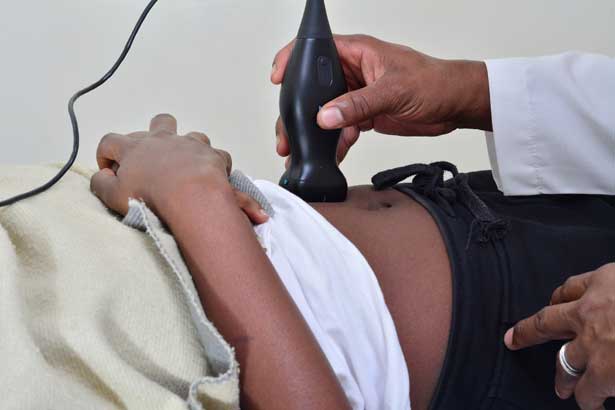
Ultrasound scans consist of a small probe known as a transducer (a small hand-held device that resembles a microphone) that is placed directly on the skin. A gel is applied to the skin before placing the transducer over it. High-frequency sound waves produced in the device move from the transducer through the gel into the body. Sound is then absorbed by the tissues and reflected back to the transducer. The sound waves are converted to electrical impulses that are interpreted as black and white images.
Read on to learn more about the ultrasound scan services offered at Limuru Cottage Hospital.
Table of Contents
- Obstetrics Ultrasound Scans
- General ultrasound
- How to prepare for an obstetric ultrasound
- Frequently asked questions about ultrasound scans
- Are ultrasound scans only for pregnancies?
- How often will I get Ultrasound scans during pregnancy?
- Can too many Ultrasound scans harm the baby?
- Can babies hear ultrasounds?
- Can an Ultrasound scan result in deafness?
- Do Ultrasound scans have side effects?
- Can Ultrasound scans damage internal organs?
- Why would a doctor order an abdominal ultrasound scan?
- Can Ultrasound scans lie about the weeks of pregnancy?
Our commitment through our Ultrasound services is to provide you with the highest quality Ultrasound scan in an environment that makes the experience both enjoyable and safe for the mother to be, and unborn child.
Our Ultrasound services include:
- Obstetrics Ultrasound scans
- General Ultrasound scans
Obstetrics Ultrasound Scans
An Obstetrics Ultrasound scan is the use of sound waves that are used to create real-time visual images of the growing fetus and pregnant uterus in the womb. It is done every three to four weeks throughout the pregnancy. Under the Obstetrics Ultrasound services, we have the following scans and screenings:
Dating ultrasound scans:
Since most pregnant women are usually unsure of their “Last Monthly Period” (LMP), the dating scan, therefore, helps in accurately availing this information in regards to the pregnant mother’s due date. This scan also finds out whether you are having more than one baby (multiple pregnancies; twins, triplets, etc.).
First-trimester obstetrics ultrasound scans:
Here, an Ultrasound scan is first done to ascertain the viability of a pregnancy. This Ultrasound service gives a risk assessment of your baby’s health and development as well as giving a risk assessment for Down syndrome. It may be done either trans-abdominal (the transducer is placed over the abdomen) or trans-vaginally (where the transducer is narrow and placed in the vagina). Sometimes, both may be done simultaneously.
It is done before 13-14 weeks into the pregnancy. Our specialists will talk you through the entire scan while adequately keeping you informed on what they are looking at. Should the pregnant mother or couple have any concerns or questions, the sonographer should be in a position to address them appropriately.
Second and third-trimester obstetric ultrasound scans:
These are scans done to help assess fetal growth and anatomy. This may also include evaluation of the fetal position, amniotic fluid volume around the fetus in the uterus, location of the placenta, fetus’s cardiac activity, the fetal spine, and any fetal anomalies.
Anatomy Ultrasound scans:
This is important to monitor the development and growth of your baby, including the heart (looking for holes and ensuring the vessels are positioned correctly), adequate brain development, spine, nose, lips for cleft palate, arms and legs, etc.
Gender scans:
Gender scans are obstetric Ultrasound services usually done at 15 weeks into the pregnancy. The scan is brief where the pregnant mother to be is shown the baby. The gender can be shown to you at the time of the scan or placed in an envelope for you to do a gender reveal at the time of your choosing.
General ultrasound
General Ultrasound scans use sound waves to produce images of the inside of the body. It helps diagnose the causes of pain, swelling and infection in the body’s internal organs and examines the state of a fetus in pregnant women.
General Ultrasound services cover areas such as the abdominal, pelvic, trans-vaginal region, testicular/testis, breasts, and thyroid, which are all reported by your consulting radiologist. special Ultrasound scan method that evaluates the movement of materials in the body.
This scan plays an important role in spotting kidney defects, muscle and tendon tears, gall stones, fatty liver disease, cysts, liver tumours etc.
A Doppler Ultrasound scan may be part of the Ultrasound services. Doppler scan is a special Ultrasound scan method that evaluates the movement of materials in the body. It enables the doctor to see and evaluate blood flow through the arteries and veins in the body. There are three common types of Doppler ultrasound:
- Color Doppler; It uses a computer to convert Doppler measurements into a collection of colours to show the speed and direction of blood flow through a vessel.
- Special Doppler; This one shows blood flow measurements graphically, in terms of the distance travelled against a unit of time, instead of as a colour picture. It can also convert blood flow information into a unique sound that can be heard with every heartbeat.
- Power Doppler; This is a superior technique that is more sensitive than colour Doppler and is capable of giving better detail of blood flow, especially when the blood flow is little or minimal. It however does not help the radiologist determine the direction of blood flow, which may be important in some cases.
Vascular ultrasound:
Vascular ultrasounds are a type of scan often used to test for such things as carotid arteries, varicose veins and leg arteries. These will subsequently enable your doctor to treat vessel narrowing and prevent things like ulcers etc.
You are welcome to contact us at any time to help you monitor and nurture your fetus into a healthy baby while taking care of your health as a mother through the different Ultrasound services available in our facilities.
How to prepare for an obstetric ultrasound
The preparation needed for a trans-abdominal or trans-vaginal Ultrasound scan is minimal. When an obstetric Ultrasound scan is being performed after 18 weeks of pregnancy, some doctors will advise their patients to arrive with a full bladder. If a transvaginal Ultrasound is to be performed, the patients are advised to empty the bladder by urination just before the Ultrasound scan is done. This reduces discomfort and relaxes the bladder for a better view of the pelvic organs.
Ultrasound transducers are not disposable hence, it is advisable for patients to visit a medical centre that follows proper hygiene protocols because they need proper cleaning before and after each patient’s use to avoid infections. Trans-abdominal Ultrasound transducers are cleaned with disposable antiseptic wipes while transvaginal transducers are usually covered with a disposable cover during the scan.
Since most Ultrasound scans are painless and fast, after the scan is done, the patient can return home right after and continue with their usual activities.
Frequently asked questions about ultrasound scans
Are ultrasound scans only for pregnancies?
Fetal imaging is one of the most common uses of ultrasounds services, but not limited to it so it’s just one of the several applications of Ultrasound services.
How often will I get Ultrasound scans during pregnancy?
Most healthy women will receive two Ultrasound scans throughout the pregnancy. The first during the first trimester to confirm the due date, and the second at 18-22 weeks to confirm the normal anatomy of and sex of the baby.
Can too many Ultrasound scans harm the baby?
Having too many Ultrasound scans during pregnancy is unlikely to cause any long-lasting harm to the growing fetus.
Can babies hear ultrasounds?
Fetuses can hear ultrasounds during scans and the sound is as loud as a subway train entering the station.
Can an Ultrasound scan result in deafness?
With protracted exposure during Ultrasound scan services, an inaudible scan can also result in hearing loss.
Do Ultrasound scans have side effects?
Internal and external Ultrasound scans are generally painless and don’t have any known side effects apart from a little discomfort that may result from a probe being pressed over your skin or inserted into your body.
Can Ultrasound scans damage internal organs?
There is no known case of adverse reactions from the performance of ultrasounds in human beings.
Why would a doctor order an abdominal ultrasound scan?
Your doctor may recommend that you have an abdominal Ultrasound scan if you’re at risk of an abdominal aortic aneurysm. An abdominal Ultrasound scan can help your doctor determine the cause of stomach pain or bloating, as well as check for kidney stones, liver disease, tumours, and many other conditions.
Can Ultrasound scans lie about the weeks of pregnancy?
Ultrasounds are known to more accurately predict your due date than using your last menstrual period, though only in the first trimester and early second trimester (until roughly 20 weeks into the pregnancy).